Human Embryo Image Segmentation
Developed an end-to-end pipeline for robust segmentation of 2-cell-stage human embryos in grayscale microscopic images using advanced computer vision techniques.
Project Overview
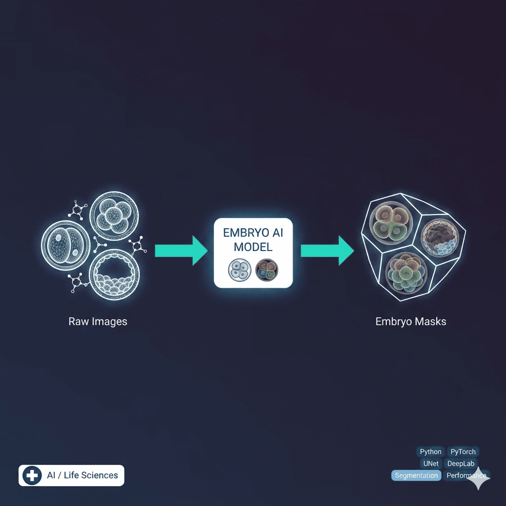
Presented a comprehensive end-to-end pipeline that incorporates all considerations for the robust segmentation of a 2-cell-stage human embryo in grayscale microscopic images. The primary processing tools include gamma correction, feathered circular masking, and bandpass filtering in frequency space, which enhance structural detail. Implemented Otsu thresholding and watershed segmentation with extended minima to account for positional drifts of the blastomere and signature non-uniformities, thereby effectively separating the two embryonic cells. The method demonstrates adaptability to other embryo images with slight manual tuning, making it valuable for reproductive medicine and embryology research.
Key Features
- ✓End-to-end embryo segmentation pipeline
- ✓Gamma correction for image enhancement
- ✓Feathered circular masking technique
- ✓Bandpass filtering in frequency space
- ✓Otsu thresholding implementation
- ✓Watershed segmentation with extended minima
- ✓Blastomere positional drift compensation
- ✓Non-uniformity signature handling
- ✓Two embryonic cell separation
- ✓Adaptable to various embryo images
- ✓Medical imaging processing pipeline
- ✓Robust structural detail enhancement
Technical Challenges
- ⚡Handling grayscale microscopic image variations
- ⚡Accounting for positional drifts in blastomeres
- ⚡Managing signature non-uniformities in embryo images
- ⚡Developing robust segmentation for medical applications
- ⚡Ensuring accuracy in embryonic cell separation
- ⚡Creating adaptable pipeline for different embryo stages
Technologies Used
Project Info
Screenshots